 If you’re anything like me, you spend a significant portion of the day wondering about the paths viruses take when they’re cruising around your internals. Luckily for us, a newly developed microscope from Duke researchers can show the exact path taken by the little critters (?), down to the micrometer.
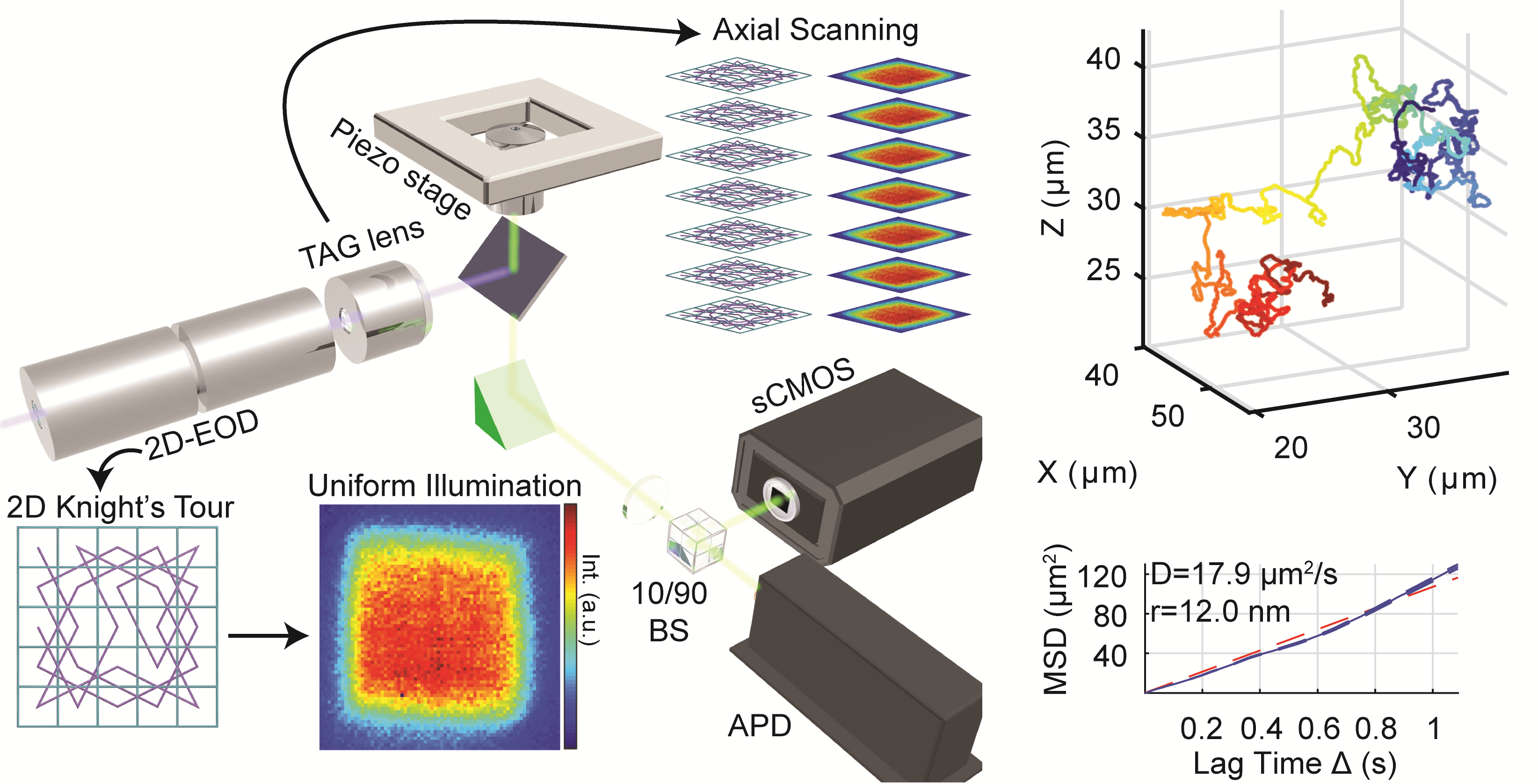
The system, designed by a team led by assistant professor Kevin Welsher, isn’t like a traditional microscope. Instead of magnifying an image using natural or augmented light, it scans a laser through a small volume repeatedly and from multiple angles. This illuminates special fluorescent particles, the positions of which can be tracked over time.
Attach one of those particles to something else and you can track what it’s doing. It’s kind of like a mocap studio for microbiology. But until recently, those particles were too big to attach to viruses — imagine trying to do your Gollum impression with basketballs taped all over your body. Welsher’s team recently improved the power of the system enough that it can detect much smaller dots — and even fluorescent proteins built right into the virus’s system. The result, as you see up top, is quite a detailed little track!
Simple, right?
I’m reminded of the old Family Circus cartoons, with Billy or whoever going all over the neighborhood, petting dogs, tracking mud on the neighbor’s porch and so on. Except Billy is a lentivirus, and the neighborhood is the soupy exterior of a cell membrane.
It’s not all just for kicks, of course: The goal is to be able to watch as a virus makes contact with a cell and does whatever it does to penetrate and infect it. That moment, so critical to understanding viral behavior, is poorly understood because it’s been nearly impossible to observe directly.
“What we are trying to investigate is the very first contacts of the virus with the cell surface — how it calls receptors, and how it sheds its envelope,” said Welsher in a Duke news release. “We want to watch that process in real time, and to do that, we need to be able to lock on to the virus right from the first moment.”
With this system, we’re a step closer to understanding one of the most sophisticated biological machines ever created. The team’s work is published this week in the journal of the Optical Society.